Areas Of The Back Anatomy / 1 / The lumbar and sacrum region make up the bone of the lower back anatomy.. These sections are cervical (neck), thoracic (upper and middle back), lumbar (lower back), and sacrum (tailbone). The back of house is the area in which food is stored and prepared, and it typically includes other staff areas such as a break room and changing area. Bones of the pelvis and lower back. Anatomy of the back organs. Each vertebra consists of the following parts:
The anatomy of the hip and back is comprised of numerous parts that can be injured or wear out, and many problems that occur in this area can display the exact same symptoms or pathology. Anatomy of the spine overview. The back's structure is complex. The vertebral column consists of 33 vertebrae which can be split up into 5 continuous sections. The spine is made of 33 individual bones stacked one on top of the other.
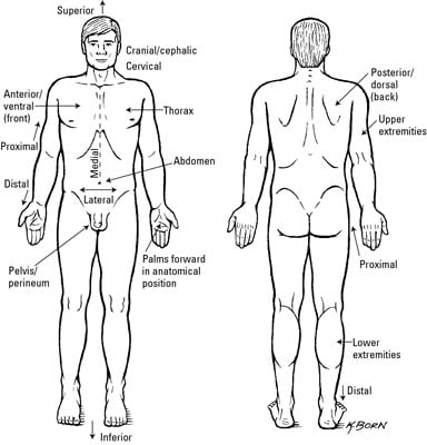
Clinical Anatomy Terms To Describe The Eight Body Regions Dummies from www.dummies.com Familiarize yourself with the parts of your back and why they are causing you pain. Anatomy of the back organs. This is my video about the muscles of the back. The back anatomy includes the latissimus dorsi, trapezius, erector spinae, rhomboid, and the teres major. The vertebrae in the lumbar spine area are the largest of the entire spine, so the lumbar spinal canal is larger than in the cervical or thoracic parts of the spine. The outer portion contains neurons, and the inner area communicates with the cerebral cortex. The bones of the pelvis and lower back work together to support the body's weight, anchor the abdominal and hip muscles, and protect the delicate vital organs of the vertebral and abdominopelvic cavities. The back is found posteriorly and includes the vertebral column, the muscles that support the back and the spinal cord.
The middle back consists of the spine (spinal column), spinal cord, nerves, discs, muscles, blood vessels, ligaments, and tendons.
These structures work together to support the body, enable a range of movements, and send messages from the brain to the. Back muscles anatomy here include the trapezius, latissimus dorsi, rhomboid and levator scapulae. The vertebral column consists of 33 vertebrae which can be split up into 5 continuous sections. The outer portion contains neurons, and the inner area communicates with the cerebral cortex. The back of house is the area in which food is stored and prepared, and it typically includes other staff areas such as a break room and changing area. The back anatomy includes the latissimus dorsi, trapezius, erector spinae, rhomboid, and the teres major. Back anatomy the back is the body region between the neck and the gluteal regions. First, we're going to look at the bone structures. The vertebral column runs the length of the back and creates a central area of recession. The spine's four sections, from top to bottom, are the cervical (neck), thoracic (abdomen,) lumbar (lower back), and sacral (toward tailbone). Lumbar nerves (5 pairs) feeds the lower back and legs. Familiarize yourself with the parts of your back and why they are causing you pain. Throughout the spine, intervertebral discs made of.
The lumbar region of the spine, more commonly known as the lower back, is situated between the thoracic, or chest, region of the spine, and the sacrum. To put it plainly, sometimes hip pain comes from the hip, but a lot of times hip pain comes from the back. Surface anatomy of the back. Areas on the skin surface supplied by nerve fibers from one spinal root. The back consists of the spine, spinal cord, muscles, ligaments, and nerves.
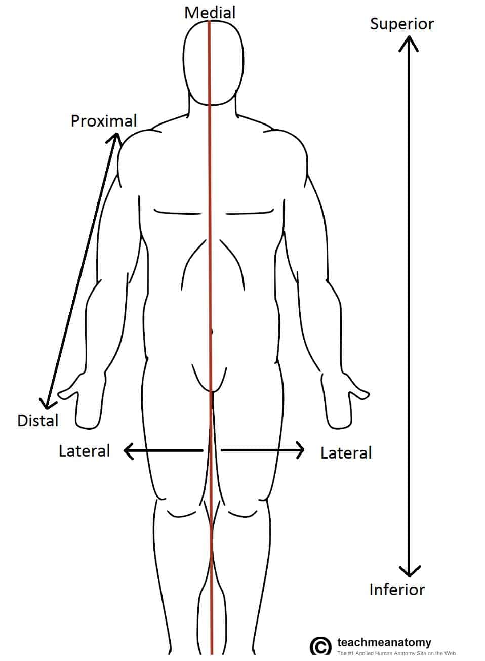
Anatomical Terms Of Location Anterior Posterior Teachmeanatomy from teachmeanatomy.info On this page, you'll learn about each of these muscles, their locations and functional anatomy. Surface anatomy of the back. Bones of the pelvis and lower back. The anatomy of the hip and back is comprised of numerous parts that can be injured or wear out, and many problems that occur in this area can display the exact same symptoms or pathology. Back muscles anatomy here include the trapezius, latissimus dorsi, rhomboid and levator scapulae. First, we're going to look at the bone structures. To put it plainly, sometimes hip pain comes from the hip, but a lot of times hip pain comes from the back. Because of its size, the lumbar spine has more space for the nerves to move about.
The back anatomy includes the latissimus dorsi, trapezius, erector spinae, rhomboid, and the teres major.
Human body anatomy female female anatomy muscle shoulder blade pain anatomy back muscles bones man female anatomy body muscles in a body female anatomy muscole shoulder concept muscular sysyem. You have 33 vertebrae (bones) that make up the vertebral column. The bones of the pelvis and lower back work together to support the body's weight, anchor the abdominal and hip muscles, and protect the delicate vital organs of the vertebral and abdominopelvic cavities. The spine's four sections, from top to bottom, are the cervical (neck), thoracic (abdomen,) lumbar (lower back), and sacral (toward tailbone). The human back, also called the dorsum, is the large posterior area of the human body, rising from the top of the buttocks to the back of the neck. The back's structure is complex. The outer portion contains neurons, and the inner area communicates with the cerebral cortex. Then we may think of little else. There are a variety of anatomical structures that make up the anatomy of the foot and ankle (figure 1) including bones, joints, ligaments, muscles, tendons, and nerves. Lumbar nerves (5 pairs) feeds the lower back and legs. Each vertebra consists of the following parts: The vertebral column consists of 33 vertebrae which can be split up into 5 continuous sections. The spine runs from the base of your skull down the length of your back, going all the way down to your pelvis.
There are 5 areas covered in the upper limb: It comprises the vertebral column (spine) and two compartments of back muscles; The 5 vertebrae in the lower back form the lumbar region of the spine. The axilla is a pyramidal shaped anatomical area which gives passage for neurovascular structures, such as the brachial plexus and axillary vein, to enter and leave the upper limb. Each vertebra consists of the following parts:
Back Anatomy All About The Back Muscles from www.kingofthegym.com This is my video about the muscles of the back. With a good grasp of foot anatomy it readily becomes apparent which surgical approaches can be used to access various areas of the foot and ankle. First, we're going to look at the bone structures. The back of house is the area in which food is stored and prepared, and it typically includes other staff areas such as a break room and changing area. These structures work together to support the body, enable a range of movements, and send messages from the brain to the. The cervical region is the most flexible region of the spine, followed by the lumbar region and the thoracic region. As you can see from the image below, your back, or spine, is made up of many parts. Human body anatomy female female anatomy muscle shoulder blade pain anatomy back muscles bones man female anatomy body muscles in a body female anatomy muscole shoulder concept muscular sysyem.
To put it plainly, sometimes hip pain comes from the hip, but a lot of times hip pain comes from the back.
The back is found posteriorly and includes the vertebral column, the muscles that support the back and the spinal cord. It comprises the vertebral column (spine) and two compartments of back muscles; Anatomy of the back organs. The axilla is a pyramidal shaped anatomical area which gives passage for neurovascular structures, such as the brachial plexus and axillary vein, to enter and leave the upper limb. Connects portions of the upper abdomen and muscles in the back and chest areas. The anatomy of the back refers to the muscles of the back, as well as the bones of the scapulae, ribcage, and spine. The human back, also called the dorsum, is the large posterior area of the human body, rising from the top of the buttocks to the back of the neck. The spine runs from the base of your skull down the length of your back, going all the way down to your pelvis. These structures work together to support the body, enable a range of movements, and send messages from the brain to the. These sections are cervical (neck), thoracic (upper and middle back), lumbar (lower back), and sacrum (tailbone). Familiarize yourself with the parts of your back and why they are causing you pain. There are a variety of anatomical structures that make up the anatomy of the foot and ankle (figure 1) including bones, joints, ligaments, muscles, tendons, and nerves. It is the surface of the body opposite from the chest and the abdomen.
The axilla, cubital fossa, extensor tendon compartments, carpal tunnel and anatomical snuffbox areas of the back. Low back pain is a very common complaint for a simple reason.


0 Komentar